
If you have been referred for a pelvic or abdominal ultrasound, you probably have questions about what to eat and drink beforehand, what the scan actually looks at, and why your doctor chose ultrasound over other options like MRI or CT. This article covers all that in plain language, so you can arrive prepared and know what to expect.
What is an abdominal or pelvic ultrasound?
An ultrasound is a scan that uses high-frequency sound waves to create real-time images of the structures inside your body. It does not use radiation. A handheld device called a transducer is moved gently across the skin. It sends sound waves into the body and records the echoes that return. A computer converts those echoes into the images your sonographer and radiologist review.
Abdominal ultrasound looks at the organs in your abdomen. It includes the liver, gallbladder, spleen, pancreas, kidneys, and large blood vessels in this area. Pelvic ultrasound focuses on the organs in the lower abdomen or pelvis. In women, this typically includes the uterus, ovaries, and surrounding structures; in men, it may include the bladder and prostate.
Because ultrasound produces live images and does not require radiation, it is one of the most widely used medical imaging modalities in Australia. According to the Australian Institute of Health and Welfare, more than 27 million Medicare-subsidised diagnostic imaging services were performed in Australia in 2022 to 2023 - across X-ray, ultrasound, CT, MRI, and nuclear medicine combined (AIHW, Diagnostic Imaging Services, 2022-23).
How do you prepare for an abdominal ultrasound?
Preparation for an abdominal ultrasound is straightforward. Simply following the instructions carefully helps your sonographer get the clearest possible images for your radiologist to assess.
Fast for four to six hours before your appointment. This means no food and no drinks other than water during this period. Fasting lowers gas in your digestive system. It also lets the gallbladder fill with bile and this makes it easier to see on ultrasound. If you need to take regular medications, you may take these with a small sip of water - confirm this with your doctor if you are unsure.
Water consumption is not typically recommended unless required for medications. Please consult your doctor or Qscan Radiology clinic prior to your scan if unsure.
You do not need a full bladder for a standard abdominal ultrasound unless your referral also includes a pelvic component, in which case the preparation instructions in the next section apply.
A typical abdominal ultrasound appointment takes around 20 to 45 minutes, depending on the number of structures being assessed and whether additional views are needed.
How do you prepare for a pelvic ultrasound?
Preparation for a pelvic ultrasound depends on the type of scan your doctor has requested. If age appropriate, we always offer transabdominal and transvaginal scans as part of the routine examination, which will require full bladder preparation.
Transabdominal pelvic ultrasound
For a transabdominal pelvic ultrasound - where the transducer is placed on the skin of the lower abdomen - you will need a full bladder. A full bladder acts as an acoustic window, pushing loops of bowel out of the way and allowing clear views of the uterus and ovaries underneath.
To achieve this, drink about one litre of water in the hour before your appointment. Do not empty your bladder until after the scan. Your Qscan team will confirm the exact timing and fluid amount when you book, as this can vary slightly depending on the structures being assessed.
Transvaginal ultrasound
A transvaginal ultrasound is a type of pelvic ultrasound in which a small, smooth probe is gently inserted into the vagina. This approach provides a closer, more detailed view of the uterus and ovaries and is often used alongside or instead of the transabdominal approach. For this type of scan, an empty bladder is preferred - you will be asked to use the bathroom before the scan begins.
Your sonographer will explain the procedure before it starts and will only proceed with your consent. If you have any concerns or questions, please tell the team before or during your appointment.
Combined abdominal and pelvic ultrasound
If your referral covers both the abdomen and pelvis, preparation will require both fasting and filling for optimal visualisation. The team will advise you clearly when you book.
Quick reference: preparation by scan type.
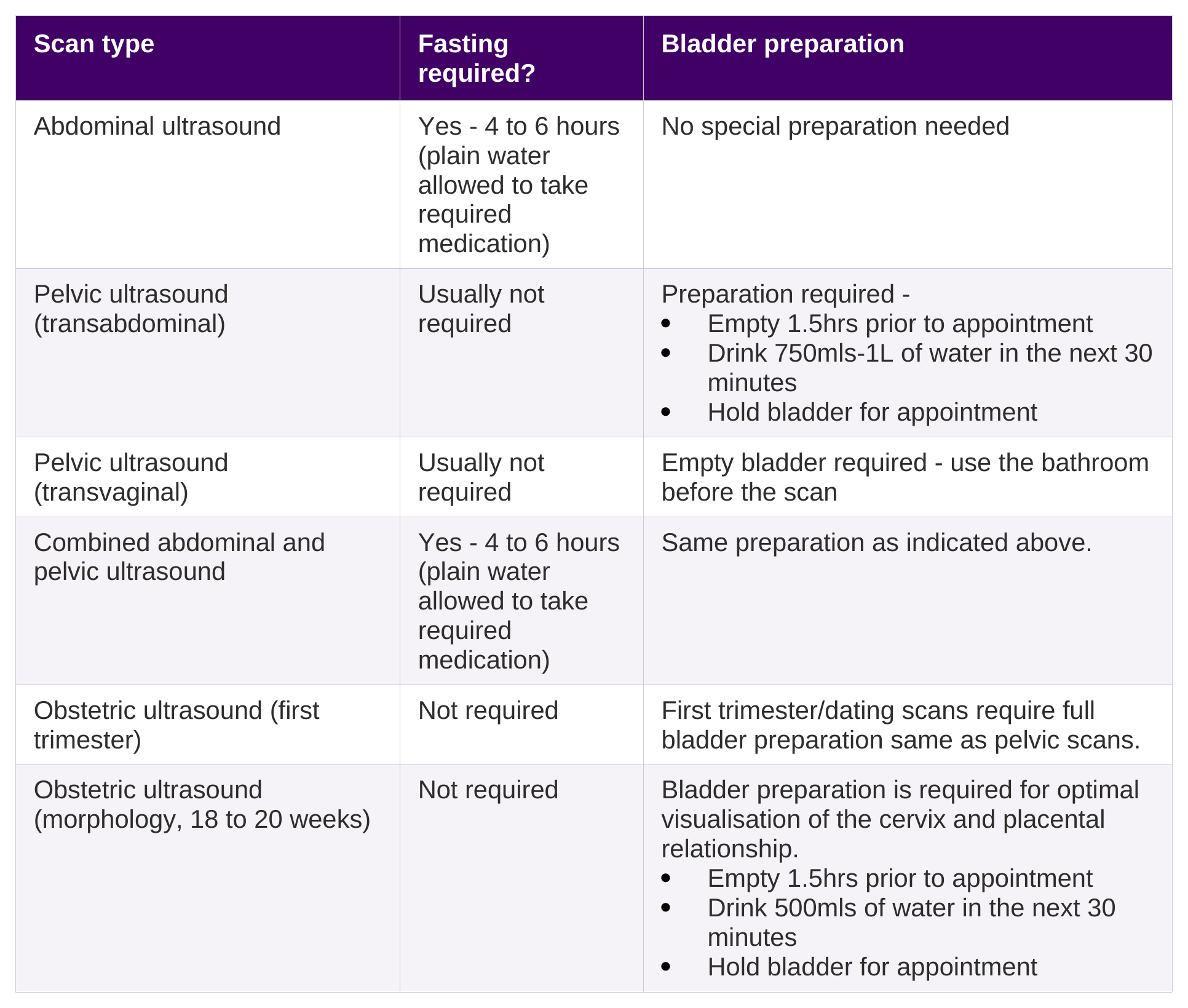
If you are unsure about your preparation instructions, call your Qscan clinic or check your referral. Incorrect preparation is one of the most common reasons a scan needs to be rescheduled.
What does an abdominal ultrasound show?
An abdominal ultrasound gives your doctor a detailed view of the soft tissue organs in your abdomen. Particularly well suited to assessing:
Liver
-
To assess size and texture
-
Investigate abnormal liver function tests
-
Check for fatty liver, cysts, or masses
Gallbladder and bile ducts
- To look for gallstones or inflammation
- Assess causes of abdominal pain, particularly after eating
- Evaluate bile duct size if blockage is suspected
Pancreas
- To investigate upper abdominal pain
- Assess for inflammation or structural changes (where visible)
Spleen
- To check for enlargement
- Investigate infections, blood disorders, or trauma
Kidneys
- To assess kidney size and structure
- Investigate pain, infection, or reduced kidney function
- Check for kidney stones or obstruction
Aorta and major blood vessels
- To screen for abdominal aortic aneurysm (AAA)
- Assess blood flow and vessel size
Free fluid within the abdomen
- To detect fluid that may be related to infection, inflammation, or other medical conditions
Ultrasound is often the first imaging test doctors use for abdominal symptoms. It does not use radiation. It is widely available. It gives real-time information about an organs structure and its associated characteristics. For some findings, your doctor may request a follow-up scan using MRI or CT to get additional detail - this is a normal part of the imaging pathway and not necessarily a cause for concern.
What does a pelvic ultrasound show?
A pelvic ultrasound helps your doctor assess the organs and structures in the pelvis. What is assessed depends on your sex, your symptoms, and what your referring doctor has asked for. In females, the scan focuses on the female reproductive organs, while a male pelvis examination is more commonly performed as a renal tract scan, covering the bladder and prostate.
In women, pelvic ultrasound is commonly used to assess:
Uterus
-
To assess size, shape, and position
-
Investigate abnormal bleeding or pelvic pain
-
Check for fibroids, polyps, or other structural changes
Endometrium (lining of the uterus)
- To measure thickness and appearance
- Investigate irregular or postmenopausal bleeding
- Monitor changes related to the menstrual cycle or hormone therapy
Ovaries
- To assess size and appearance
- Investigate pelvic pain or hormonal symptoms
- Check for cysts, follicles, or masses
Fallopian tubes (where visible)
- To look for fluid or swelling that may suggest blockage or infection
Cervix
- To assess structure and identify any abnormalities if visible
Bladder
- To evaluate bladder filling and emptying
- Investigate urinary symptoms if relevant
Free fluid within the pelvis
- To detect fluid that may be related to ovulation, inflammation, or other conditions
According to the Royal Australian and New Zealand College of Obstetricians and Gynaecologists (RANZCOG), transvaginal ultrasound is recommended as the preferred first-line imaging investigation for suspected endometriosis (RANZCOG, Endometriosis Clinical Practice Guideline, 2024-25). At Qscan, we also offer dedicated 'Deep Infiltrating Endometriosis' (DIE) assessments at select locations. This is a separate scan from our standard pelvic ultrasound, requiring a longer appointment time and a higher fee.
In men, renal (pelvic) ultrasound may be used to assess:
Bladder
- To assess bladder shape and wall thickness
- Investigate symptoms such as frequency, urgency, or discomfort
- Check for stones, masses, or other abnormalities
Bladder emptying (post-void residual)
- To measure how well the bladder empties after urination
- Investigate incomplete emptying or urinary retention
Prostate gland
- To assess size and appearance
- Investigate urinary symptoms such as weak stream or difficulty starting urination
- Check for enlargement or other changes
Seminal vesicles
- To assess structure where visible
- Investigate certain reproductive or prostate-related concerns
Distal ureters (where visible)
- To look for dilation that may suggest obstruction
Free fluid within the pelvis
- To detect fluid that may indicate inflammation, infection, or other underlying conditions
Ultrasound, MRI, or CT: which scan does your doctor choose and why?
Your referring doctor selects the most appropriate imaging modality based on the clinical question they are trying to answer, your individual circumstances, and the strengths of each modality. The table below explains how the three main modalities compare for abdominal and pelvic assessment.
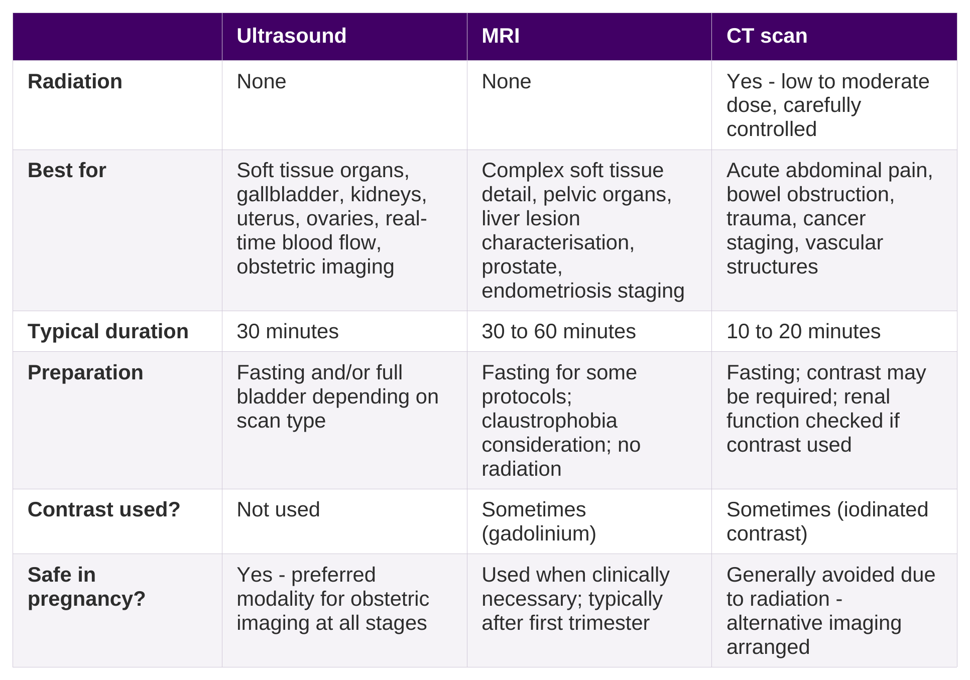
The right scan depends on the clinical question, not on which modality is more advanced. A gallstone, for example, is reliably assessed on ultrasound and does not routinely require MRI. A complex liver lesion identified on ultrasound may need MRI for further characterisation. Your referring doctor makes this decision based on your history, symptoms, and what they need to know.
When would your doctor choose MRI over ultrasound for the pelvis or abdomen?
MRI is often used when a more detailed assessment of soft tissue is needed, or when ultrasound findings require further characterisation. Common reasons your doctor might request an MRI after or instead of an ultrasound include:
-
Suspected deep infiltrating endometriosis - when transvaginal ultrasound is unavailable or when deep endometriosis is suspected, pelvic MRI may be offered to assess the extent of changes (RANZCOG, Endometriosis Clinical Practice Guideline, 2024-25)
-
Liver lesion characterisation - MRI with liver-specific sequences can help characterise lesions identified on ultrasound
-
Prostate assessment - multiparametric MRI is widely used as the recommended investigation for clinically suspected prostate cancer (RANZCR)
-
Uterine fibroid assessment prior to treatment planning
-
Staging of known or suspected gynaecological or gastrointestinal malignancy
Ultrasound and MRI are often complementary - one does not replace the other. It is common for a patient to have an ultrasound first and for MRI to follow if the initial findings require further investigation.
Booking your abdominal or pelvic ultrasound at Qscan
Qscan sonographers perform abdominal and pelvic ultrasounds across our clinic network in Queensland, New South Wales, ACT, Tasmania, and Western Australia. A referral from your GP or specialist is required.
To book, find your nearest Qscan location or simply book online. When you book, the team will confirm your preparation instructions so you arrive ready for your appointment.
For more information about ultrasound at Qscan, visit our Ultrasound page, our Pelvic Ultrasound page, or our Abdominal Ultrasound page.
Frequently Asked Questions
Why do I need a full bladder for a pelvic ultrasound?
A full bladder creates an acoustic window - it pushes loops of bowel out of the way and allows the ultrasound waves to travel clearly to the uterus and ovaries underneath. Without a full bladder, the bowel can obstruct the view and make it difficult for your sonographer to see the pelvic organs clearly. This is only needed for the transabdominal approach. For a transvaginal ultrasound, an empty bladder is required.
Can I eat before a pelvic ultrasound?
For a pelvic ultrasound on its own, fasting is usually not required. If your scan also covers the abdomen - particularly the gallbladder, liver, or kidneys - you will need to fast for four to six hours beforehand. Check your referral or confirm with the Qscan team when you book to make sure you have the right preparation instructions for your specific scan.
What is the difference between a transabdominal and a transvaginal ultrasound?
A transabdominal ultrasound places the probe on the skin surface of the lower abdomen. A transvaginal ultrasound uses a narrow internal probe for a closer view of the uterus and ovaries. Your sonographer or referring doctor will advise which approach is needed. In many cases, both techniques are used in the same appointment to provide the most complete picture.
Does a pelvic or abdominal ultrasound hurt?
Most patients find an abdominal ultrasound comfortable. The gel applied to the skin may feel cool, and gentle pressure from the transducer is normal. For a transvaginal ultrasound, some patients experience mild discomfort from the probe, particularly if there is underlying pelvic tenderness. Tell the sonographer if you experience anything uncomfortable during the scan so they can adjust.
How long does it take to get results?
Your radiologist's report is typically sent to your referring doctor, and available via the Qscan Patient Results app, around 7 to 10 days after your scan. Your referring doctor will then discuss the results with you and advise on next steps.
Can I have an ultrasound if I am pregnant?
Yes. Ultrasound is the standard imaging modality for obstetric assessment and is used safely throughout all stages of pregnancy. If you are pregnant and have been referred for a non-obstetric ultrasound, let the team know when you book so they can confirm the most appropriate approach for your situation.
|
This article is general information only and is not a substitute for medical advice. Your individual circumstances may differ. Speak with your doctor about whether imaging is right for you. Bulk billing applies to eligible Medicare-rebatable services and is subject to MBS item number, patient eligibility, and clinic. Out-of-pocket costs may apply. Our team will confirm any costs with you when you book. |